Lameness exams
Lameness accounts for more losses in money and time than any other condition in the equine industry. There are widely varying degrees of lameness, ranging from subtle reduced performance to complete loss of mobility requiring euthanasia.
 Understanding the basics of lameness, and working with our team using a lameness exam and diagnostic techniques, can help get your horse back on his feet.
Causes of Lameness
Lameness arises from a musculoskeletal injury or defect in the leg. This includes wounds to skin, connective tissue bruising, muscle pain, arthritis (joint inflammation), tendon sheath and bursa inflammation, tendon and ligament injury, and injuries to bone.Some lameness is a result of conformation of the limb, while other lameness is a direct effect of external influences to the body. Most front end lameness is typically caused by a structure in the foot or ankle, while most hind end lameness is caused by a problem in the hock or stifle. Isolating the source of the lameness requires the expertise of a skilled veterinarian.
Diagnosis
A thorough lameness exam is the cornerstone of lameness diagnosis. It is a detailed veterinary procedure and includes the following steps.:
 1. A complete history, including when the lameness first occurred, the work program the horse is currently in, and details about the horse including breed and age.
2. A thorough standing exam including evaluation of conformation, hooftester exam, and palpation of the joints and soft tissues of the limbs.
3. Examination of the horse in movement, either in hand or under saddle, to evaluate the horses’s gait in straight lines and on circles, at the walk, trot and canter. The degree of lameness is assessed before flexion tests are performed.
4. Flexion exams, stressing specific joints or regions of the limb for a specified time. The limb is held in flexion, the horse trotted off, and the lameness compared to the baseline degree of lameness. This helps identify the source of the lameness. As with many parts of the exam, flexion tests are interpreted in consideration of what is normal for that specific horse.
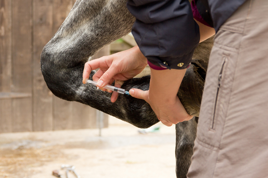
5. If the lameness is not easily isolated during the flexion tests, nerve blocks are often used. Nerve blocks are used to methodically numb portions of the limb. A temporary “block” is produced with injection of a local anesthetic around specific nerves or into specific joints or other structures. The horse is assessed at the trot before the block. Then the area in question is numbed, and the horse is asked to trot off again. If there is no improvement, the process is continued on specific nerves, progressing up the limb until the lameness is lessened or gone. This identifies the specific region of the pain
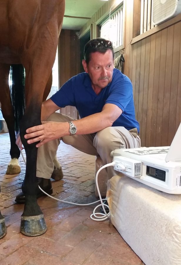
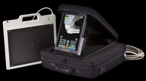 6. Once the area of interest is identified, diagnostic imaging can then be used to visualize the structures in the area. Imaging may include x-ray of bone, ultrasound of soft tissues, or less common modalities like magnetic resonance imaging (MRI), computerized axial tomography (CAT scan) and nuclear scintigraphy (two-dimensional imaging).
The complete lameness exam is a very important tool to identify performance problems in the horse. Carried out and interpreted correctly, the lameness exam should provide an accurate diagnosis. The usefulness of the lameness exam relies heavily on the expertise of the veterinarian. To be performed effectively, it requires a thorough understanding of anatomy, hands-on experience, and a methodical approach. Because some of these component tests are subjective, a lameness exam is as much an art as it is a science.
Lameness Treatments
Treatment of lameness highly depends on the cause. Once a thorough lameness exam and diagnostic imaging has been performed, we can move on to treating the cause.
Examples of treatments include:
- Confined stall rest or a period of turnout to rest injured structures.
- Joint (intra-articular) injections of steroids and other substances to reduce inflammation and pain in a joint.
- Oral or injectable anti-inflammatories and pain relievers to manage multiple problems, to manage chronic pain in older or debilitated horses, and as an adjunct to more specific therapies used.
- Surgery (performed in a hospital setting) is sometimes used to treat certain types of lameness. The most common is arthroscopic surgery, wherein repairs are made to the joint surface through several tiny portals and using a tiny camera and instruments inserted into the joint.
- There are now a multitude of other novel therapies available including Extra-Corporeal Shockwave Therapy, PRP Therapy, IRAP Therapy and others. These are generally classed as “regenerative therapies” wherein the body is manipulated in some way to heal itself without externally derived medications.